Muscles Are Activated in the Pupillary Light Reflex.
Light is the stimulus. Which muscles are activated when the pupil responds to light.

Central Control Anatomy And Physiology I
Gastroileal reflex is a reflexstimulated by the opening of the ileocecal valve pushing the digested material from the ileum.

. Solution for Which muscles are activated when the pupil responds to light. First week only 499. Smooth muscles are activated in the pupillary light reflex.
Afferent limb of reflex is the optic nerve. Effector- smooth muscle of iris. Smooth muscles are activated in the pupillary light reflex.
What muscles are activated in the pupillary light reflex. And the response is conveyed to the pupillary musculature by autonomic nerves that supply the eye. What is the specific cellular receptor involved in the pupillary reflexes.
In medicine there are two pupillary reflexes-. Testing the pupillary light reflex is easy to do and requires few tools. The pupillary light reflex two main parts.
Vision is not necessary for the reflex to approximation and there are no clinical conditions in which the light reflex is present but the reflex to the approximation is. Rod and cone cells. At the same time the IML also receives excitation from the LC which is itself excited by the.
The pattern of pupillary response to light can help determine which of. This reflex is also called the inverse myotatic reflex because it is the inverse of the stretch reflex. Reset Help Skeletal The nervous system predominately controls the pupillary light reflex.
Smooth muscles are activated in the pupillary light reflex. Which part is the effector of the reflex action. Images displayed onto the retina are upside-down and backward.
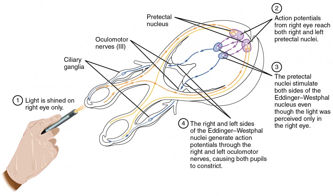
Parasympathetic The pupillary light reflex is an example of an Somatic reflex Sympathetic Autonomic reflex. First sensory connects each retina with both pretectal nuclei in the midbrain at the level of the superior colliculi. The reflex to approximation synkinesia not the true reflex is activated when looking from a distant object to a close one.
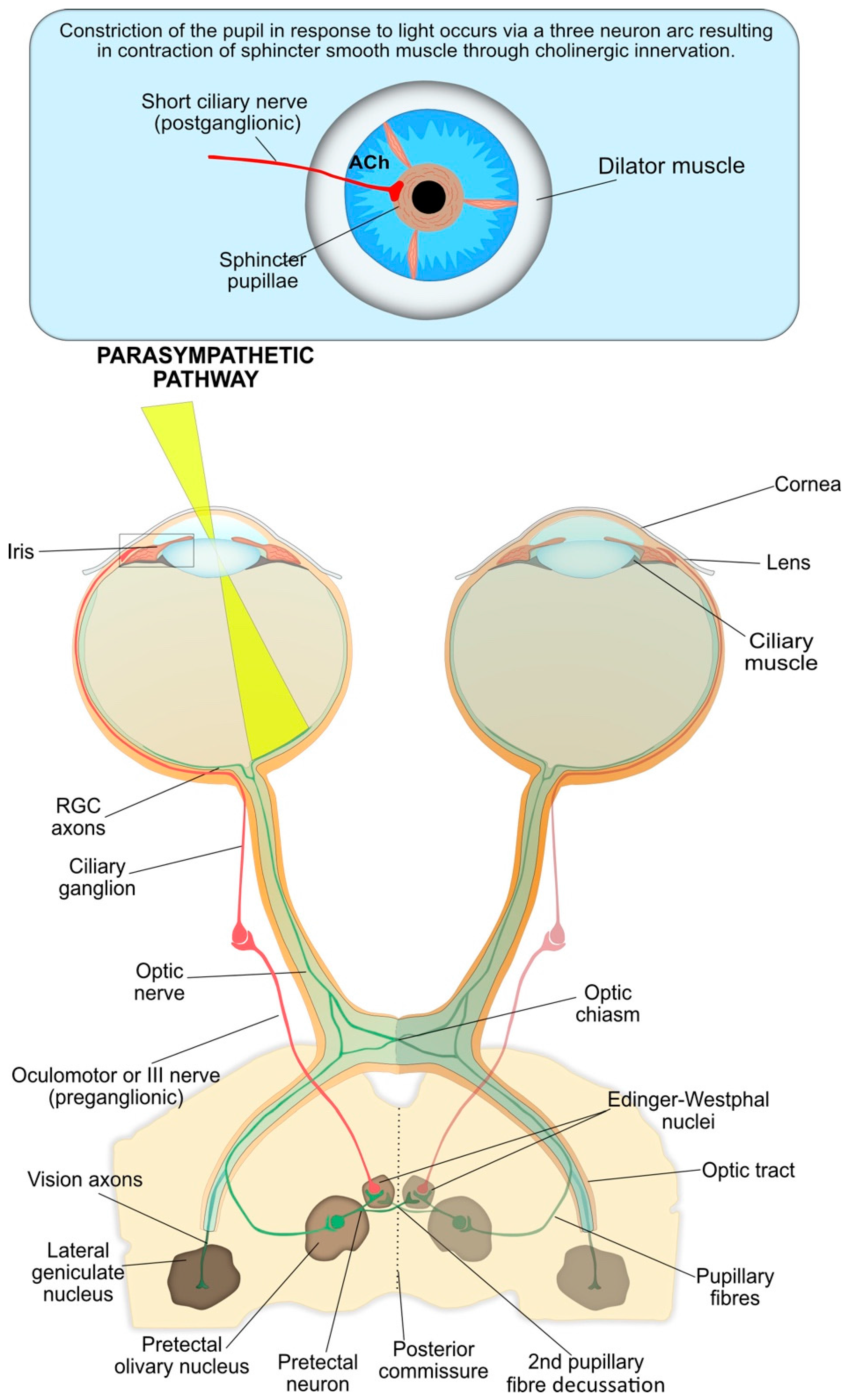
The diagram below shows the neuroanatomical pathways. Drag the labels to identify the five basic components of the pupillary light reflex pathway. This learning objective details the pupillary light reflex which allows for the constriction of the pupil when exposed to bright light.
Up to 24 cash back The pupil reflex is the reflex contraction and relaxation of the antagonistic muscles of the iris in response to changes in light intensity. Efferent pathway- oculomotor nerve. The pupillary accommodation reflex is the reduction of pupil size in response to an object coming close to the eye.
Nucleus SCN which activates the pupillodilator muscle PD causing pupil dilation. Includes accommodation convergence and miosis. Ø Pupillary fibers travel along with the other fibres transmitting through the optic nerve.
Start your trial now. The pupil of the right eye constricts while shining a flashlight into the left eye. Stimulation of parasympathetic fibers in the oculomotor nerve III causes contraction of the sphincter pupillae muscles muscarinic.
The pupil dilates at low light intensities and constricts at high light intensities. The primary components of the reflex arc are the sensory neurons or receptors that receive stimulation and in turn connect to other nerve cells that activate muscle cells or effectors which perform the reflex action. Both these reflexes affect both eyes even if only one eye is stimulated.
Images displayed onto the retina are upside-down and backward. It causes a change in the pupil size. Smooth muscles are activated in the pupillary light reflex.
Smooth muscles are activated in the pupillary light reflex. This reflex serves to regulate the amount of light the retina receives under varying illuminations. However antagonistic muscles are activated.
You just studied 7 terms. An afferent limb and an efferent limb. Click card to see definition.
The pupil either constricts or dilates to respond to light and near stimuli. Although muscle tension is increasing during the contraction the alpha motor neurons in the spinal cord that supply the muscle are inhibited. The pupillary light reflex PLR is the constriction of the pupil that is elicited by an increase in illumination of the retina.
The size of our pupils changes continuously in response to variations in ambient light levels a process known as the pupillary light reflex PLR. Tap again to see term. While there are other reasons for variation in pupillary dilation and constriction such as arousal leading to changes in the balance of the sympathetic and parasympathetic nervous systems here we will focus on.
The direct PLR present in virtually all vertebrates is the constriction of the pupil in the same eye as that stimulated with light. Light entering the eye is processed through the pupillary light reflex and signals directed to the iris sphincter muscle to adjust the amount of light that reaches the retina. Which of the following statements does not describe the procedure for testing the pupillary light reflex as shown in the video.
What are the 5 parts of the reflex arc of the pupillary light and consensual reflex. What is the main stimulus for the pupillary light reflex. While there are other reasons for variation in pupillary dilation and constriction such as arousal leading to changes in the balance of the sympathetic and parasympathetic nervous systems here we.
Light entering the eye is processed through the pupillary light reflex and signals directed to the iris sphincter muscle to adjust the amount of light that reaches the retina. The pupillary light reflex is an example of an Autonomic reflex. Now up your study game with Learn mode.
The pupillary sphincter and dilator muscles are the two involuntary iris muscles that are required to control the amount of light traveling to the retina. Muscle Light reflex - The light reflex is mediated by the retinal photoreceptors and subserved by four neurones A. The pupillary light reflex is the reduction of pupil size in response to light.
The iris sphincter regulates the amount of light entering the eye to keep it within the dynamic range of the photoreceptors. The consensual PLR is the constriction of the pupil in the eye opposite to the eye stimulated with light. The PLR is not a simple reflex as its function is modulated by cognitive brain function and any long-term.
The pupillary sphincter and dilator muscles are the two involuntary iris muscles that are required to control the amount of light traveling to the retina. Impulses reach the brain via the optic nerve. The pupillary light reflex allows the eye to adjust the amount of light that reaches the retina.
Click again to see term. MIOSIS constriction is mediated via the PUPILLARY LIGHT REFLEX. Afferent pathway- optic nerve.
Tap card to see definition.

Pupil Light Reflex Pathway Red And Blue Lines Represent The Afferent Download Scientific Diagram

Pupil Light Reflex Pathway Red And Blue Lines Represent The Afferent Download Scientific Diagram

Diagnostics Free Full Text Eyeing Up The Future Of The Pupillary Light Reflex In Neurodiagnostics Html
0 Response to "Muscles Are Activated in the Pupillary Light Reflex."
Post a Comment